Filter
Exclude
Time range
-
Near
ntusbs
Registration and abstract submission are now open for the Singapore Bioscience Symposium (SBS) 2026! 🗓️ 7–9 December 2026 📍NTU Singapore Learn more and register: ntu.edu.sg/sbs/symposium/sbs… #SBS2026 #NTUsg #CellBiology #StructuralBiology
1
1
214
BiologyAIDaily
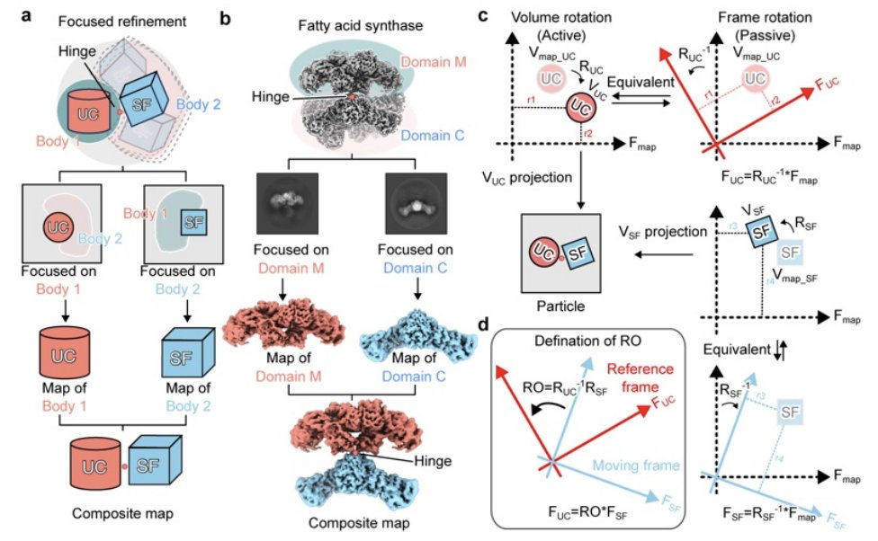
CryoROLE: Describing large inter-domain rotation in single particle cryo-EM 1. CryoROLE is a lightweight, math-based tool to recover continuous inter-domain motions that are otherwise “lost” when cryo-EM workflows produce high-resolution composite maps via focused refinement of separate rigid bodies. 2. Key idea: each particle already has multiple pose assignments (one per focused-refined domain). CryoROLE computes per-particle relative orientation (RO) between domains directly from these orientations, making the motion observable without deep learning or PCA. 3. The output is an RO “landscape”: a point cloud where each point is a particle and its coordinates encode the relative rotation between domains. Local point density reports how populated particular poses are, enabling an intuitive view of motion range preferred orientations. 4. The method is designed for large rigid-body-like motions where consensus-map-based continuous-heterogeneity methods (e.g., latent-space or perturbation models) struggle, and where classic classification discretizes a continuum into a few bins. 5. Implementation details that matter in practice: CryoROLE does statistics in rotation-vector space (avoids Euler-angle singularities), visualizes in fixed-axis Euler space for interpretability, and can “canonicalize” axes so α/β/γ align with primary/secondary/tertiary motion directions. 6. Validation on human fatty acid synthase (hFASN): with ~2.2M particles, the RO landscape accurately predicts reconstructions from particles sampled at chosen landscape coordinates, confirming that landscape coordinates correspond to real inter-wing orientations. 7. INO80–hexasome application: merging particles from 3 previously classified states reveals a continuous ~110° trajectory of INO80 rotating around the hexasome, with multiple preferred orientations. CryoROLE recovers a low-occupancy state placing the ATPase near SHL −1 that was missed by discrete classification. 8. Ribosome application (thermo-annealing): by computing RO landscapes for LSU vs SSU-body (ratchet), SSU-head vs SSU-body (swivel), and SSU-head vs LSU, CryoROLE shows annealing contracts the motion distributions and shifts population toward a preferred, low-motion basin. 9. Functional anchoring: published translocation-related 70S structures map onto the RO landscape along a plausible trajectory; particle density differences across mapped states suggest the landscape approximates thermodynamic preference. Additionally, E-site tRNA-bound particles occupy a narrower RO region, indicating tRNA restricts global ribosome motions. 💻Code: github.com/yifancheng-ucsf/c… 📜Paper: biorxiv.org/content/10.64898… #cryoEM #structuralbiology #computationalbiology #proteinDynamics #singleParticleAnalysis #openSource #bioinformatics
3
9
1,550
BiologyAIDaily
TCR-FramePose: A local-frame representation for decomposing global docking and CDR3 loop geometry in TCR-pMHC recognition 1. The paper introduces TCR-FramePose, a geometry descriptor that decomposes TCR–pMHC pose into three pMHC-referenced rigid bodies: the whole TCR variable domain, CDR3α, and CDR3β—so global docking and CDR3-local pose can be compared in the same coordinate system. 2. For each body, pose is split into reach (distance), offset direction (unit vector on S²), and orientation (rotation on SO(3), represented via quaternions). This preserves the natural separation between translation and rotation, which conventional descriptors often mix. 3. For statistics and ML, FramePose maps the manifold variables to Euclidean tangent coordinates at Fréchet means: 1 reach 2 offset 3 orientation coordinates per body (6 total), yielding 18 coordinates per complex. Orientation axes are interpretable as groove-axis roll, cross-groove pitch, and groove-normal twist. 4. On 378 curated αβ TCR–pMHC crystal structures (282 class I, 96 class II), FramePose recovers known class-associated placement differences (similar to centroid-based TCR-CoM) and additionally resolves class-associated orientation shifts (whole TCR and especially CDR3β) that crossing angle fails to capture. 5. The same orientation coordinates identify noncanonical docking modes: a tight “reverse-polarity” cluster dominated by ~170° groove-normal twist (spanning both class I and class II examples), plus distinct off-axis ~180° flip modes that look similar in magnitude but differ in axis composition. 6. In cross-validated association modeling, FramePose predicts buried surface area (BSA) well when using all three bodies (best configuration), and shows that beyond global placement, CDR3-local orientation contributes additional BSA-associated signal. 7. For binding affinity (Kd-based strong vs weak; n=244), rigid-body pose alone is only modestly informative overall, but FramePose improves over conventional docking descriptors. The best-performing configuration uses CDR3α CDR3β (not whole-TCR), indicating the affinity-associated signal is concentrated in CDR3-local geometry. 8. Feature attribution and augmentation analyses localize the most nonredundant signal to CDR3β orientation (and secondarily CDR3α orientation and CDR3β offset). These are also the least “recoverable” from conventional descriptors, explaining why they add new information. 9. Biological determinant analysis (conditioned PERMANOVA on native manifold distances) suggests docking geometry is organized primarily by germline V-region framework. After controlling for antigen context and germline framework, CDR3 sequence does not detectably reposition rigid-body pose, while MHC allele and peptide length contribute smaller, localized adjustments—especially in CDR3β and groove-normal orientation axes. 10. Interface interpretation: across the affinity-annotated cohort, affinity correlates most strongly with interface burial (BSA). CDR3β reach provides a geometric readout of burial (greater reach → reduced BSA), and the reach–affinity association attenuates after adjusting for BSA/shape complementarity, supporting a burial-linked (not independent) pose relationship. Within engineered peptide panels, mutation-level pose/contact effects are panel-specific, with CDR3β remodeling recurring in a similar interface region but varying in direction by receptor. 📜Paper: biorxiv.org/content/10.64898… #TCR #immunology #structuralbiology #computationalbiology #pMHC #proteinstructure #geometricdeeplearning #bioinformatics #Tcell #cancerimmunotherapy
7
29
2,497
BiologyAIDaily
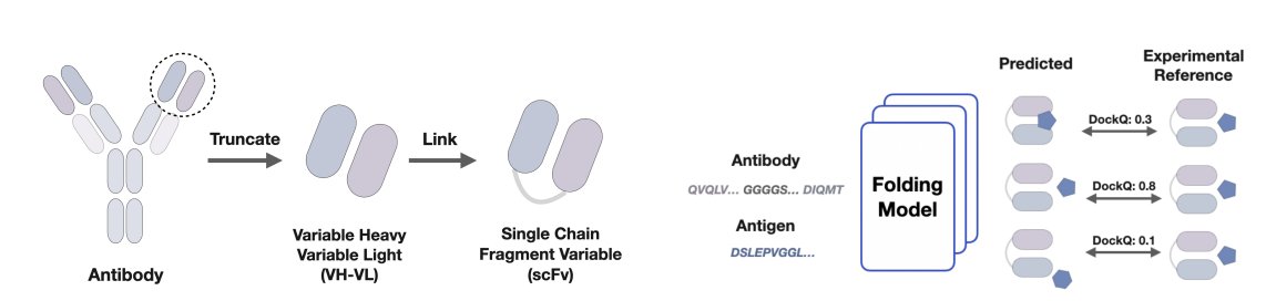
Folding scFv–Antigen Complexes at Scale 1. The paper introduces SCALE (scFv–Ag CompLex Ensembles): a large benchmarking resource to stress-test modern cofolding models specifically on scFv–antigen docking, where correct epitope engagement and binding geometry are often the true failure mode. 2. SCALE is built from a curated subset of 3,800 experimentally solved Ab–Ag complexes from SAbDab, standardized by converting each heavy/light antibody into an scFv sequence (VH (GGGGS)3 linker VL) paired with a single antigen chain, then deduplicated by scFv–antigen sequence identity. 3. Using multiple state-of-the-art folding backbones (AlphaFold 2.3 Multimer, AlphaFold 3, Boltz-2, Chai-1, Pairmixer) and diverse inference-time settings (random seeds, recycling depth, optional antibody MSA, optional templates), the authors generate ~197,900 predicted scFv–Ag complexes—an ensemble-centric view rather than single-shot evaluation. 4. Interface accuracy is evaluated with DockQ against experimental references, separately for VH–Ag and VL–Ag interfaces (and often summarized as their mean). Across ~200k predictions, most DockQ scores are very low; near-native interfaces (DockQ > 0.8) exist but are rare for every model, indicating that correct docking is not consistently recovered even when tertiary folds look confident. 5. Best-of-ensemble behavior is notably better than the typical sample: with enough trials, many targets yield at least one “acceptable” interface (DockQ > 0.23). Still, 879/3,800 complexes never exceed DockQ 0.23 under any tested setting, while only 256 exceed 0.23 across all conditions—highlighting a large “hard target” regime. 6. The study finds strong coupling between VH–Ag and VL–Ag interface quality (Pearson r = 0.958), with VL–Ag slightly harder on average. This suggests that when docking fails, it often fails globally (wrong pose/epitope) rather than in only one variable domain. 7. A central result: confidence metrics commonly used in binder pipelines (ipTM, ipSAE, pDockQ, pDockQ2, AbEpiScore) correlate well with DockQ when pooling all predictions globally, but perform poorly at selecting the best structure within each target’s ensemble (low per-complex correlations and low Top-1 accuracy). In other words, they separate “easy vs hard targets” better than “best vs second-best pose for the same target.” 8. The paper documents a key practical pitfall: high single-chain confidence does not imply correct complex formation. Many predictions have high pLDDT yet extremely low DockQ, underscoring a decoupling between confident tertiary structure and correct quaternary docking. 9. Inference-time choices matter but mainly for best-case outcomes: more recycling can occasionally refine already-good predictions toward near-native interfaces without shifting the median much; additional seed sampling often shows diminishing returns, but a subset of targets benefits substantially. Including an antibody MSA improves the frequency of higher-quality interfaces even though antibody sequences may have limited evolutionary signal, while templates provide only modest gains in this setting. 10. Physics-based interface descriptors computed with PyRosetta (e.g., shape complementarity, estimated binding energy, clash/repulsion terms) correlate with DockQ roughly as well as learned confidence scores for ranking, suggesting that reranking/scoring—possibly combining model confidences with physical interaction features—is a major bottleneck for scalable scFv–Ag docking. 💻Code: huggingface.co/datasets/ravi… 📜Paper: biorxiv.org/content/10.64898… #ComputationalBiology #ProteinFolding #Antibodies #StructuralBiology #Benchmark #AlphaFold #Docking #MachineLearning #Bioinformatics
6
28
2,192
febos41
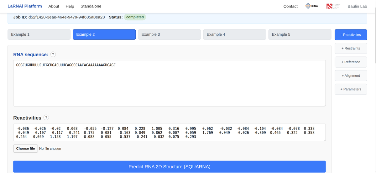
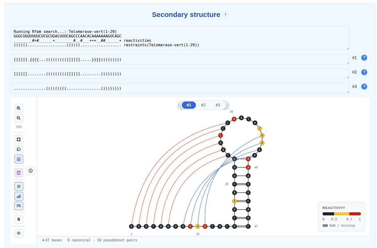
We’ve just released a new SQUARNA preprint: doi.org/10.64898/2026.06.30.… Alongside it, we are launching the LaRNAl Platform: larnal.imol.institute/ #RNA #SQUARNA #LaRNAl #LaRNAlP #2D #Structure #Prediction #Bioinformatics #StructuralBiology #ComputationalBiology #Algorithms
1
2
11
1,439
WLA_summit
Structural biology is facing a critical limitation: most atomic structures are determined from isolated, purified proteins frozen in artificial states. #Nobelist Kurt Wüthrich outlined the roadmap to break this boundary by shifting structural biology in vivo – directly inside living cells and higher organisms. While current cryo-electron tomography offers low resolution, Wüthrich championed the untapped potential of combining Nuclear Magnetic Resonance (NMR) spectroscopy with Magnetic Resonance Imaging (MRI), fusing high-resolution molecular interaction data with precise geographic anatomical mapping. Watch his full keynote speech: youtube.com/watch?v=7tVffwkH… #StructuralBiology #Biology #WLS #WLS26 #Science #BasicScience #Dubai #UAE
22
WLA_summit
For decades, structural #biologists deduced molecular dynamics by using chemical blockers to halt reactions – a method #Nobel Laureate Joachim Frank candidly calls "cheating". Speaking at the 2026 #WLS, Frank unveiled a disruptive solution: integration of a PDMS-based microfluidic chip with gas-assisted sprayers. By mixing components within half a millisecond and instantly vitrifying the product on a falling grid, Frank's lab mapped the real-time, 10-millisecond trajectory of ribosome subunit association, elevating cryo-EM from a static imaging tool to a high-speed video camera for the nano-world. Watch his full speech at the World Laureates Summit: youtube.com/watch?v=F0YdObvv… #CryoEM #StructuralBiology #Biology #WLS26 #Science #BasicScience #Dubai #UAE
1
2
37
BiologyAIDaily
CryoACE: An Atom-centric Framework for Accurate and Automated Model Building in Cryo-EM 1. CryoACE is presented as an end-to-end, atom-centric generative framework that builds full atomic graphs directly from cryo-EM density maps, targeting both homogeneous (single-structure) and heterogeneous (dynamic ensemble) reconstructions. 2. The central architectural shift is atom-centric reconstruction: instead of relying on expensive voxel-wise 3D convolutions as the main pathway, CryoACE samples density features directly at predicted atomic coordinates (via trilinear interpolation) and uses these “atomic profiles” to refine coordinates. 3. This design enables an iterative atomic self-refinement loop at inference: the model first predicts a coarse structure from sequence density, then re-samples density at the newly predicted atom positions to create better local features, repeating for multiple refinement cycles. 4. CryoACE integrates three modalities during training: (i) sequence/MSA features encoded with a Boltz-1-style MSA module Pairformer, (ii) density map features encoded from 3D patches with a 3D ResUNet plus 3D rotary positional embeddings, and (iii) atom profiles as localized density descriptors tied to candidate coordinates. 5. Multimodal fusion is implemented with cross-attention where sequence tokens query density tokens, aiming to enforce sequence/evolutionary constraints while still grounding coordinates in experimental density—especially important in low-resolution or noisy regions. 6. For heterogeneity, CryoACE introduces a training-free guidance scheme during diffusion sampling with a staged schedule: early-time global guidance aligns overall topology to a density-derived point cloud (weighted k-means Sinkhorn divergence), while late-time Q-guidance refines local atomic placement using predicted Q-scores. 7. A key practical addition is direct prediction of local resolution and per-atom/residue Q-scores via lightweight heads: predicted local resolution is used as a prior to manage ambiguous heterogeneous regions, and predicted Q-scores both accelerate inference and provide signals for late-stage guidance. 8. The work also contributes a curated training dataset (10,915 density–structure–sequence triplets, <4 Å, pre-2025) with a two-stage alignment pipeline and strict filtering (including Q-score thresholds and a “structure modeled rate” criterion) to reduce map-model misalignment and incomplete-structure noise. 9. On the Cryo2StructData test set (excluded from training), CryoACE reports 100% completeness and improved geometric/density agreement versus neural baselines (ModelAngelo, CryoAtom, E3-CryoFold), achieving higher backbone/all-atom accuracy, lower RMSD, and the best Q-score among compared methods. 10. On real heterogeneous datasets (EMPIAR-10345 integrin; EMPIAR-10516 SARS-CoV-2 spike), CryoACE is reported to recover complete dynamic ensembles with strong map fit (weighted cross-correlation) and improved physical plausibility (lower clash rates, good MolProbity), while maintaining stable frame-to-frame structural consistency (PSC). 📜Paper: arxiv.org/abs/2606.31332 #CryoEM #StructuralBiology #ProteinStructure #DiffusionModels #DeepLearning #ComputationalBiology #ModelBuilding #Heterogeneity #Bioinformatics #AIforScience
3
15
1,957
IIMCB_Poland
🔝 Dr Wojciech Galej, who will join IIMCB in October to lead the Laboratory of RNA Splicing, has been elected an EMBO Member. 👉 iimcb.gov.pl/en/news-press-o… #EMBOMembers2026 #RNAbiology #StructuralBiology #LifeSciences #IIMCB Image credit: Stuart Ingham/EMBL
2
195
sigtrans_sttt
Usingstructural & functional analyses, this study reveals the molecular mechanisms of allosteric modulation in opioid receptors, offering insights for developing selective & safer opioid therapeutics. @UESTC1956 @SoutheastU #STTT #OpenAccess: doi.org/10.1038/s41392-026-0… #Opioids #PainResearch #DrugDiscovery #StructuralBiology
1
285
BiologyAIDaily
Adversarial Sequence Mutations in AlphaFold and ESMFold Reveal Nonphysical Structural Invariance, Confidence Failures, and Concerns for Protein Design 1. The study systematically stress-tests AlphaFold 3 with adversarial sequence perturbations and finds a striking “structural invariance”: predicted folds often remain essentially unchanged even after mutating up to 40% of residues (including deliberately destabilizing substitutions) or deleting up to 10% of residues. 2. The benchmark spans 200 proteins split across 4 bins (monomer/multimer × novel/similar to training set, using a 30% homology cutoff). Notably, fold invariance is comparable for “novel” proteins and proteins with training-set homologs, arguing that the effect is not limited to obvious in-distribution cases. 3. The mutation protocol is explicitly designed to be disruptive rather than evolution-like: point mutations swap residues in a maximally deleterious way and are biased toward central/core regions; deletions are also biased toward central positions. Mutations are cumulative across thresholds, enabling controlled trajectories from 5% to 70% (point) and 1% to 10% (deletions). 4. Using TM-score ≥ 0.5 as a same-fold criterion, AlphaFold 3 frequently preserves global topology far beyond what physical intuition would suggest. Local quality degrades earlier (lDDT drops faster than TM-score), implying that packing worsens while the overall fold “stays locked.” 5. Multimers reveal an important asymmetry: while global fold/topology can remain stable, interfaces are much more fragile. DockQ drops rapidly with mutation burden; by 20% point mutation, about half of multimer interfaces are already incorrect relative to the unmutated AlphaFold 3 prediction. 6. Fold-switching proteins (15 experimentally validated monomeric cases) do not “switch” in AlphaFold 3 under mutations targeted to known fold-switching regions. Predictions remain highly invariant up to ~40% mutation (average TM-score ~0.63 vs the original predicted structure), suggesting a disconnect from biologically plausible mutational responses even in small monomers. 7. The paper contrasts AlphaFold 3 with ESMFold (monomers only). ESMFold is more mutation-sensitive, especially for point mutations: structural similarity to its own unmutated prediction collapses earlier (notably between 20% and 40% mutation), whereas AlphaFold 3 declines more gradually and only “catches up” near 70% mutation. 8. The authors interpret the AlphaFold 3 vs ESMFold difference as potentially reflecting training objective/input modality: ESMFold’s single-sequence masked-language-model objective may couple sequence identity to structure more tightly, even though ESMFold is not claimed to be more accurate in absolute terms on standard benchmarks. 9. Confidence metrics emerge as a second major failure mode. When generating 5 candidate structures and selecting by internal ranking, AlphaFold 3 picks the most accurate structure only ~25% of the time (AlphaFold 2 up to ~35%). Even when selection is wrong, the penalty can be large (up to ~0.17 TM-score loss or >0.40 DockQ loss). 10. A template-availability analysis (FoldSeek search against pre-cutoff PDB) shows AlphaFold 3 confidence correlates strongly with the structural similarity of the best available pre-cutoff template, while sequence identity to that template is a weak predictor. This supports the concern that confidence can act as a proxy for training-set structural coverage rather than calibrated uncertainty about physical correctness—relevant for mutation interpretation and gradient-based protein design workflows that assume meaningful sequence-to-structure sensitivity. 📜Paper: doi.org/10.34133/csbj.0142 #AlphaFold3 #ProteinFolding #ProteinDesign #ComputationalBiology #StructuralBiology #ESMFold #DeepLearning #Bioinformatics #ProteinEngineering #Benchmarking
4
48
4,066
BiologyAIDaily
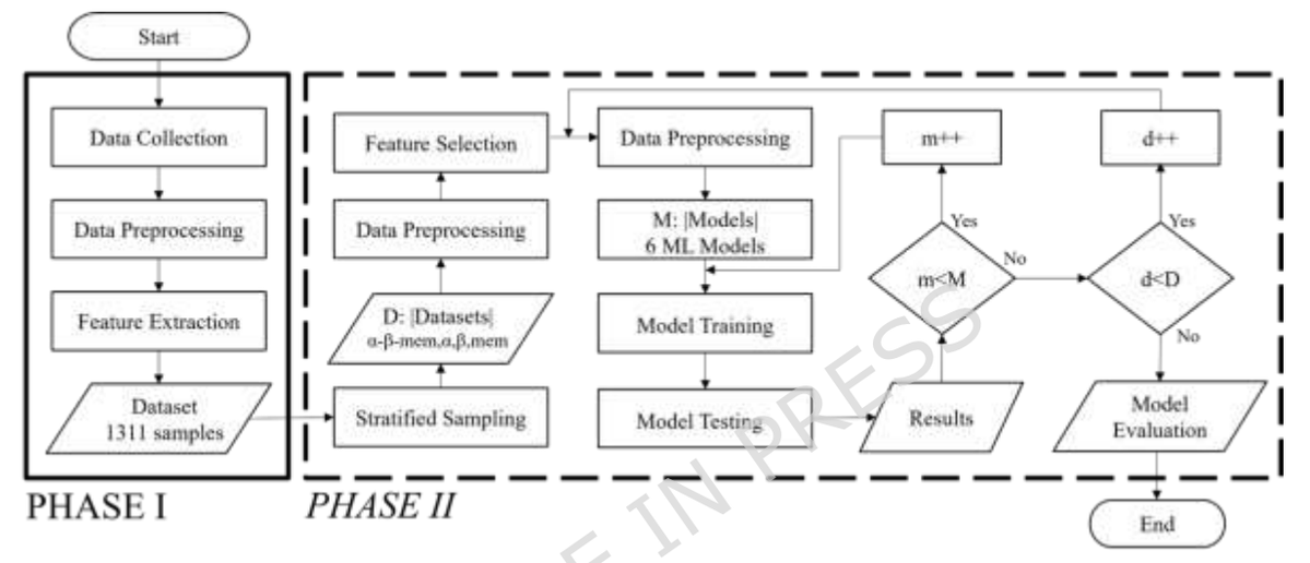
Structural feature-based machine learning benchmarking for protein interface prediction 1 This study benchmarks structure-only machine learning for residue-level protein–protein interface prediction, focusing on permanent homodimers and using features computed from unbound monomer structures (no interaction geometry required at inference time). 2 The core result: among six standard ML models (RF, XGBoost, LR, NB, MLP, KNN), Multilayer Perceptron and XGBoost performed best on a curated set of 1,311 homodimers, reaching MCC ≈ 0.94 and F1 ≈ 0.96 on the combined test setting. 3 The dataset design is a major methodological point: 1,311 non-redundant homodimer complexes (sequence similarity constrained to <85%), stratified into 422 cytoplasmic α-helical, 411 cytoplasmic β-strand, and 478 membrane complexes, enabling analysis of how interface patterns differ by secondary-structure context and environment. 4 Labels are derived from solvent accessibility changes and Levy-style region definitions: residues are categorized as core/rim/support/interface vs surface/interior, then simplified to a binary task where CORE RIM are INTERFACE and SURFACE is NON-INTERFACE (support/interior excluded as inaccessible). 5 Feature engineering starts broad (42 descriptors, mostly numerical) spanning SASA/rASA, neighborhood SASA summaries, hydrophobicity summaries, protrusion index (Cx) variants and neighborhood sums, plus multi-scale surface roughness and planarity computed on residue patches (multiple radii). 6 Interpretability and efficiency are addressed via RFECV a domain-informed refinement step: from the plateauing top features, the authors propose a compact 6-feature subset that remains highly predictive across models and categories: rASA, SASA, roughness (20 Å), planarity (20 Å), planarity (11 Å), and average protrusion index. 7 With only these six descriptors, performance stays high: on the combined α-β-Mem setting, XGBoost reaches MCC 0.912 and F1 0.94; MLP reaches MCC 0.908 and F1 0.937—showing that much of the signal is captured by a small set of biologically meaningful surface geometry/exposure features. 8 Structural stratification yields an important nuance: with full features, category-specific models are similar to the generalized model; but under the minimal 6-feature regime, category-specific training becomes more beneficial, helping recover accuracy lost from feature reduction—especially relevant for membrane proteins where interface properties are more distinct. 9 Cross-category transfer is asymmetric: α-trained and β-trained models generalize relatively well to each other, while membrane-trained models transfer worse to cytoplasmic proteins, supporting the idea that membrane interfaces occupy a different structural/environmental feature space. 10 As an external (preliminary) generalization check beyond the homodimer training domain, the approach is tested on one heterodimer (PDB 9ETL, CAPRI target). Several specialized models achieve very high scores (e.g., F1 0.985, MCC 0.981), but the paper emphasizes this is a single-case demonstration rather than broad validation. 11 The study also benchmarks against a ColabFold-based baseline for interface identification (using predicted complexes and rASA-based labeling). ColabFold shows mean accuracy ~0.90 but substantially lower mean F1 (~0.75) and MCC (~0.69) on their test distribution, and performs poorly on the 9ETL case (F1 0.298, MCC 0.156), highlighting the competitiveness of compact structure-descriptor models for this specific task setup. 📜Paper: doi.org/10.1038/s41598-026-5… #ComputationalBiology #Bioinformatics #ProteinInteractions #ProteinStructure #MachineLearning #XGBoost #NeuralNetworks #StructuralBiology #MembraneProteins #FeatureSelection
1
8
1,304
BiologyAIDaily
BoltzProt-1: Towards Efficient De Novo Binder Design with Good Developability 1 BoltzProt-1 is a de novo protein binder (incl. nanobody/VHH) design pipeline that explicitly targets two bottlenecks at once: improving prospective binding hit rates on novel targets and ensuring therapeutic-style developability (stability, solubility, low nonspecific binding, etc.). 2 The key technical shift is ranking designs with a dedicated protein–protein interaction predictor (BoltzPPI), rather than relying mainly on structure-prediction confidence proxies. In a budget-matched test on the same candidate pools, this changes selection—not generation—and directly tests whether better ranking alone improves experimental recovery. 3 On 10 low-homology (novel) targets, replacing BoltzGen’s selection with BoltzPPI ranking increases confirmed-binder rate from 3.3% (5/150) to 8.0% (12/150), a 2.4x gain. Screening hits also rise (revised hit rate 4.7% to 9.3%). Confirmed target coverage improves from 2/10 to 3/10 via ranking, and to 4/10 when paired with an improved generative model. 4 The paper also argues for stricter experimental reporting: it separates “screening hits” (including ambiguous sensorgrams) from “confirmed binders” (clean kinetics plus orthogonal confirmation). Confirmation uses a flipped assay orientation to reduce format-specific artifacts and avidity effects, with additional independent testing for some hits. 5 BoltzPPI is built on Boltz-2 representations and adds a PPI prediction head trained jointly with a confidence head. It uses interface-focused signals: token/pair features, predicted coordinates, distance embeddings, and binder/target masks, refined by a 4-block Pairformer stack (16 heads, dropout 0.25). 6 Training uses PDB and patent-derived complexes as positives, plus synthetically generated protein pairs as negatives. A multi-view training scheme drops trunk pairwise representations 50% of the time to encourage geometric reliance and reduce overfitting to internal trunk signals; additional regularization injects Gaussian noise into representations. The interaction head is trained with focal loss and combined with confidence losses. 7 On an external 10-target panel used by Chai-2, BoltzProt-1 reports screening hits on 7/10 targets, compared with 6/10 reported by Chai-2 and 3/10 for BoltzGen in this study’s setup. This suggests improved target coverage across diverse classes (signaling/adaptor proteins, cytokines/hormones, SUMOylation enzymes, calcium-binding regulators). 8 Developability is treated as a first-class outcome. Confirmed binders from the low-homology panel are evaluated across a broad assay suite (Twist Bioscience): thermal stability (Tonset, Tm1, Tm2), aggregation onset (Tagg), monomer purity (aSEC), heterogeneity (DLS PDI), hydrophobicity (HIC), polyspecificity (BVP ELISA), and self-association (AC-SINS). 9 Under combined developability criteria, 58% (7/12) of BoltzProt-1 confirmed binders pass every filter, exceeding BoltzGen confirmed binders (40%, 2/5) and clinical-stage controls measured in parallel (IgG 25%, 3/12; VHH-Fc 21%, 5/24). Attrition is minimal until hydrophobicity (HIC), which is the dominant failure point. 10 Novelty checks indicate recovered designs are not near-duplicates of known antibody/nanobody CDRs: every recovered design has minimum CDR3 edit distance ≥4 to its closest SAbDab match (with larger distances when considering CDR1 2 3). Structural context is provided via binding-site similarity to known PDB interfaces (FoldDiSCO), highlighting that the low-homology benchmark emphasizes limited prior structural precedent. 📜Paper: biorxiv.org/content/10.64898… #ComputationalBiology #ProteinDesign #DeNovoDesign #Nanobody #AntibodyEngineering #ProteinProteinInteraction #MachineLearning #DrugDiscovery #Developability #StructuralBiology
6
45
4,167
SMARTshenzhencn
SMART’s Mingxu Hu lab launches a free integrated cryo-EM data processing platform, bringing CryoSieve, CryoPROS, and other advanced algorithms into one web-based workflow. Free to use. Try it now. 🔬 #CryoEM #StructuralBiology #AIforScience #SMART
1
35
PDBeurope
🧬 New @EMBL course: "Protein Domains: Structure, Classification & Evolution" - free & online now. CATH, SCOP, Pfam, IDRs more. 🔗 ebi.ac.uk/training/online/co… #StructuralBiology #Bioinformatics #Domains
5
12
1,048
StephaneRedon
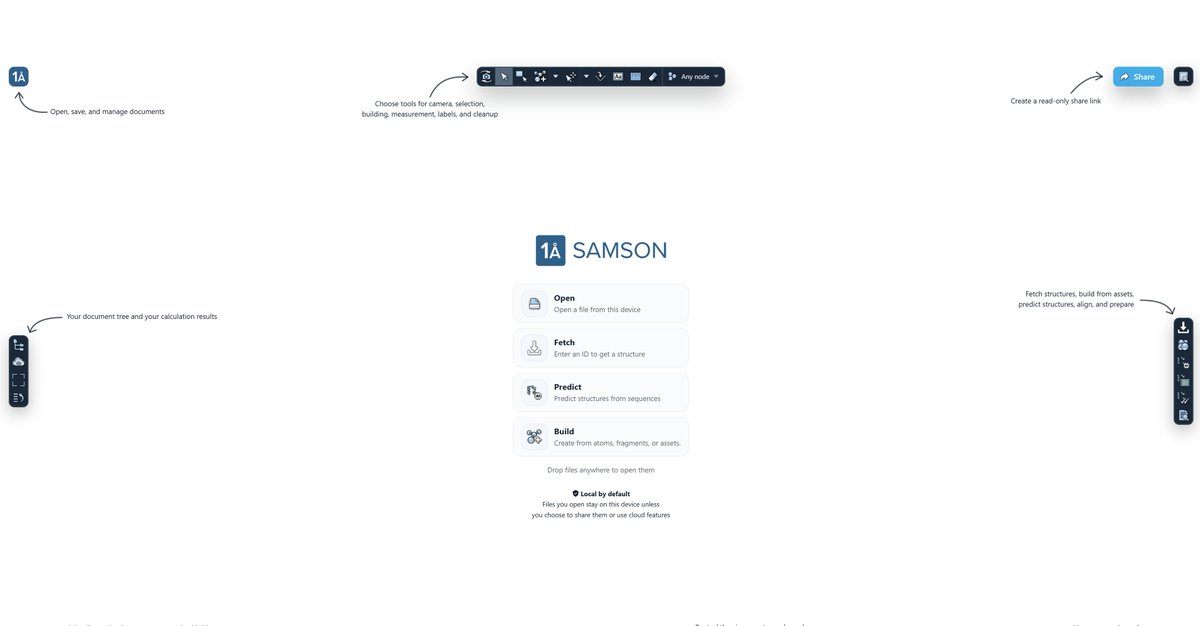
SAMSON Web is live: create, predict, render, and share molecular systems directly in your browser! Today, we are incredibly proud to introduce SAMSON Web: a browser-based molecular design environment. No installation. No plugin. No account required. Open SAMSON Web, create or import a molecular system, work on it directly in the browser, and share it. Not a screenshot. Not a video. A real interactive 3D molecular document. With the full context. With SAMSON Web, you can start in many ways: • open molecular files from your device • import and export common formats such as PDB, MOL2, and more • fetch structures from databases • predict structures (currently with AlphaFold 2, Boltz-2, and Chai-1x) • build from atoms, fragments, and assets • import cloud job results And once your system is in the browser, you can work on it: select, measure, prepare, align, minimize, annotate, edit, create a new version, and share again. Because one of the most important parts of SAMSON Web is sharing interactive documents. Scientific communication still relies too much on molecular images or movies. But molecular systems are three-dimensional. And they have context, annotations, associated data, versions, and intent. With SAMSON Web, you can share an interactive molecular document that preserves context in seconds. A collaborator, student, reviewer, customer, or colleague can open it in the browser, rotate it, zoom in, inspect annotations, associated logs and data, understand the model directly, and even edit their own copy. Remote collaborators, even during a Zoom or Teams session, can now explore designs as they want, edit, and send you back their changes. SAMSON Web also helps you create high-quality visuals directly in the browser: • advanced rendering effects • custom backgrounds • 3D skyboxes • path tracing • advanced materials • snapshots and render-ready scenes Modern molecular design produces much more than coordinates: prediction results, alignments, tables, metadata, trajectories, reports, annotations, and intermediate files. SAMSON documents preserve it all, and SAMSON Web also includes a Data Explorer to explore and edit rich scientific data next to molecular structures. The goal is to make molecular documents richer and more useful: scientific workspaces where structure, data, calculations, and interpretation live together. You start locally by default (files you open stay on your device unless you choose to share them or use cloud features). You do not need an account to create and share. Creating an account unlocks versioned documents and history, permission controls, and access to your data from anywhere. SAMSON Web is live. You can start in seconds. The link is in the first comment. #OneAngstrom #SAMSON #MolecularDesign #ComputationalBiology #DrugDiscovery #ProteinDesign #StructuralBiology #MaterialsScience #Nanoscience #AlphaFold #Boltz #Chai #MolecularVisualization #ScientificComputing #AIForScience
1
6
8
344